When It Comes to Knee Pain, The Size of Your Meniscus Matters
Elucidating why some patients with specific joint deformities are symptomatic while others are not.
Complete the form below to unlock access to ALL audio articles.
With all of the fantastic imaging techniques available in healthcare today, clinicians are capable of diagnosing tissue and joint deformities using non-invasive imaging with remarkable accuracy. However, one vexing question remains: why are some patients with specific joint deformities symptomatic while others are not?
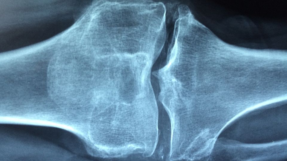
The meniscus is a piece of cartilage that cushions the knee joint between the femur (thigh bone) and the tibia (shin bone). Some people are born with a congenital morphological variation in their meniscus, called a discoid lateral meniscus (DLM), where the meniscus is thickened on the lateral, or outer side of the knee. DLM malformations cause the lateral meniscus to form a circle rather than a crescent shape, thickening the cartilage and making it more prone to tears. Some patients develop symptoms such as knee pain and a locking, leading to surgery.
Want more breaking news?
Subscribe to Technology Networks’ daily newsletter, delivering breaking science news straight to your inbox every day.
Subscribe for FREEIn order to better understand what factors separate symptomatic DLM cases from asymptomatic cases, a multi-institutional team of researchers led by Dr. Kazuya Nishino of the Graduate School of Medicine at Osaka Metropolitan University analyzed 61 knees with discoid lateral meniscus surgery without dislocation (symptomatic group) and 35 without symptoms but with discoid lateral meniscus detected on MRI (asymptomatic group). The percentage of meniscus was calculated in the coronal and sagittal sections, respectively. The researchers also measured the height of the thinnest and thickest part of the meniscus.
The results revealed that the percentage of the meniscus covering the tibia in the coronal and sagittal planes was higher in the symptomatic group than in the asymptomatic group. Additionally, the results showed that the meniscal height was greater in the symptomatic group than in the asymptomatic group.
Overall, preoperative imaging is helpful in determining the amount of tissue resection, or removal, required for patients with symptomatic DLM. “Making surgical decisions and plans based on these characteristics is expected to assist in medical treatment. In the future, we will investigate the morphological changes of the meniscus before and after surgery in three dimensions,” said Dr. Nishino.
While the difference in the morphologies of symptomatic vs. asymptomatic DLMs was the most important finding in the study, the MRIs of symptomatic patients also showed meniscal tears or other evidence of instability. The researchers suggest that these morphological features could be responsible for DLM symptoms in symptomatic patients.
Reference: Nishino K, Hashimoto Y, Kinoshita T, Iida K, Tsumoto S, Nakamura H. Comparative analysis of discoid lateral meniscus size: a distinction between symptomatic and asymptomatic cases. Knee surg sports traumatol arthrosc. 2023;31(12):5783-5790. doi: 10.1007/s00167-023-07650-2
This article has been republished from the following materials. Note: material may have been edited for length and content. For further information, please contact the cited source.