LC-MS – What Is LC-MS, LC-MS Analysis and LC-MS/MS

Complete the form below to unlock access to ALL audio articles.
From pharmaceuticals and food to bodily fluids and soil, the analytical lab has seen an increasing need for accurate measurement of microgram and sub-microgram quantities of targets, sometimes in complex matrices. While this is no mean task in itself, the potential need to analyze hundreds of samples as quickly as possible while ensuring data quality adds additional challenges.
Coupling of liquid chromatography (LC) with mass spectrometry (MS) has provided analytical scientists with a powerful tool to meet these stringent demands. Due to their versatility and efficiency, liquid chromatography-mass spectrometry (LC-MS) instruments have become desirable in many modern analytical laboratories.
What is LC-MS?
LC-MS is an analytical technique that involves physical separation of target compounds (or analytes) followed by their mass-based detection. Although relatively new, its sensitivity, selectivity and accuracy have made it a technique of choice for detecting microgram or even nanogram quantities of a variety of analytes ranging from drug metabolites, pesticides and food adulterants, to natural product extracts.
How does LC-MS work?
LC separation
LC brings about a physical separation of the analytes in a liquid sample or a solution of a solid sample. A few microliters of sample solution are injected into a flowing stream of a solvent, called the mobile phase. While the optimal injection volume is dependent on the experimental conditions, it is possible to inject as little as 0.1 µL up to as much as 100 µL of the sample accurately using an autosampler.1 The mobile phase is continuously pumped through a column (a stainless-steel tube) usually filled with silica particles coated with another liquid, the stationary phase. When the sample solution-mobile phase mix reaches the column, its components will differentially interact with the stationary phase (which remains in the column) depending upon their chemical composition or physical properties. Based on the mechanism of interaction between the analyte and the stationary phase, LC separations have been classified into different modes, such as:
- Partition chromatography – based on the differing solubility and hydrophobicity of the analytes in the stationary phase as compared to the mobile phase.
- Ion-exchange chromatography – separates the analytes on the basis of their ionic charges.
- Size-exclusion chromatography – exploits the differences in the sizes of the analyte molecules to separate them.
- Affinity chromatography – separates the analytes based on their ability to bond with the stationary phase.
Some analytes will interact more strongly with the stationary phase than others, resulting in their separation as they pass through the column. The analytes that have the least interaction with the stationary phase emerge from the column first. As the mobile phase continues to flow through the column, the remaining analytes are flushed out sequentially, those with the strongest interactions emerging last. The time a specific analyte spends in the column is characteristic of that analyte and is called its retention time (RT).
LC detection
The mobile phase flowing out of the column (the eluent) passes through a detector that “responds” to a certain physical or chemical property, such as refractive index or light absorption, of the analytes within it. This response is captured as a signal or a “peak” whose intensity (peak area or peak height) corresponds to the amount of the component present in the sample. The time at which the detector “sees” the analyte is its RT. The identity of a compound in a sample can be confirmed by comparing its RT with the RT of a known compound. While this is not an accurate method of compound identification, it helps when some information about the sample is known a priori.
Using MS for LC detection
Although a wide variety of detectors of differing technologies and sensitivities have been coupled with LC for analyzing different sample types, the mass spectrometer has emerged as a selective, sensitive and universal detector.
Unlike other detectors, the LC eluent carrying the separated analytes is not allowed to flow into the mass spectrometer. While the LC system is operated at ambient pressures, the mass spectrometer is operated under vacuum and the two are coupled through an interface. As the column eluent flows into the interface, the solvent is evaporated by applying heat and the analyte molecules are vaporized and ionized. This is a crucial step as the mass spectrometer is only capable of detecting and measuring the gas phase ions.
As the analyte ions are generated at atmospheric pressure in the interface, the process is called atmospheric pressure ionization (API) and the interface is known as the API source. Electrospray ionization (ESI) and atmospheric pressure chemical ionization (APCI) are the most commonly used sources in LC-MS analysis.
The analyte ions are drawn into the mass spectrometer where they are subjected to electric fields and/or magnetic fields. The flight paths of the ions are altered by varying the applied fields which ensures their separation from one another on the basis of their mass-to-charge (m/z) values. Post-separation, the ions can be collected and detected by a variety of mass detectors,2 of which the most common one is the electron-multiplier. When the separated ions strike the surface of the electron-multiplier (a dynode), secondary electrons are released. These secondary electrons are multiplied by cascading them through a series of dynodes. The amplified current generated by the flow of the secondary electrons is measured and correlated to the ion concentrations in the mass spectrometer at any given instant in time (Figure 1).

Figure 1: Example schematic diagram of an LC-MS setup. Credit: Cwszot, Dagui1929, CasJu, and YassineMrabet, reproduced under the Creative Commons CC0 1.0 Universal Public Domain Dedication license.
Plotting LC-MS data
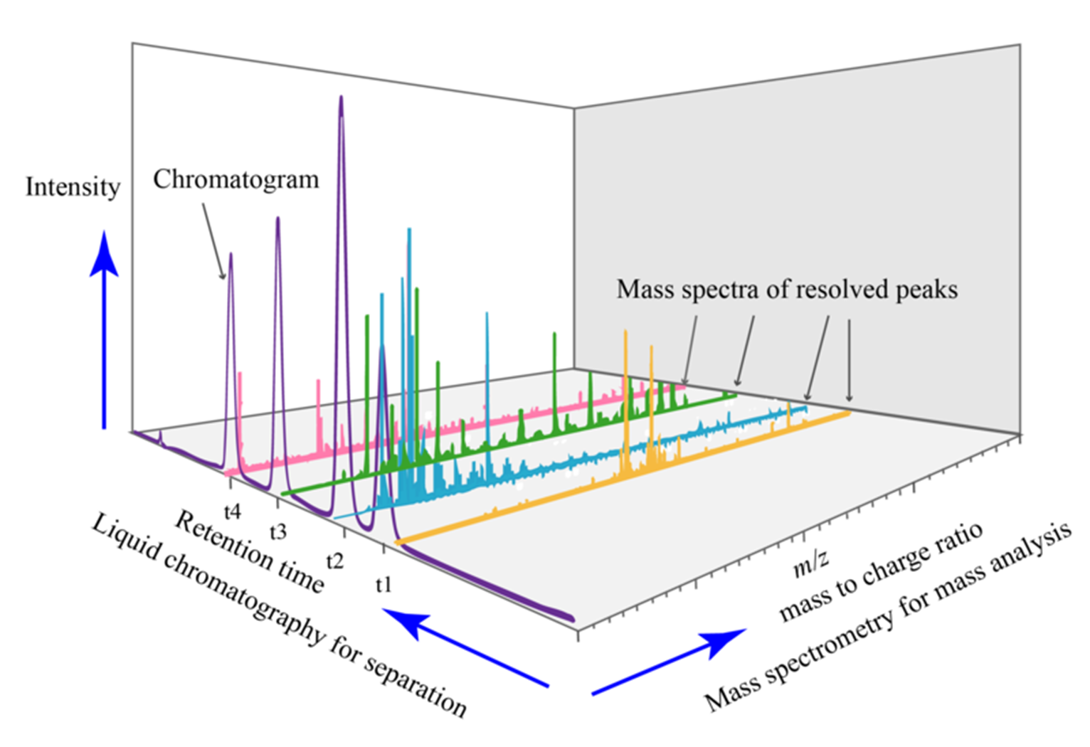
The abundances of the ions measured during the analysis of a sample by LC-MS are plotted as a total ion chromatogram (TIC). This plot displays the peak intensities of the analyte ions versus their RT. Further, each point in the chromatogram is associated with a mass spectrum. The mass spectrum depicts the ion abundances versus the measured m/z values (Figure 2).

Figure 2: Example output plot from LC-MS analysis. Credit: Daniel Norena-Caro, reproduced under the Creative Commons CC0 1.0 Universal Public Domain Dedication license.
The mass spectrum of a compound not only provides information about the mass of the parent compound (from the m/z value of its ion), but also helps to elucidate the structure of the compound from the relative abundances of isotopic mass peaks. The area of the analyte peak is used for its quantification.
The mass spectrometer can be operated in two modes, a) scan and b) selected ion monitoring (SIM). In the scan mode, it is set to detect all the ions from low m/z to high m/z values within a specified time period. This mode is used when analyzing unknown samples or when there is no available information about the ions present in a sample. When operating in SIM mode, the mass spectrometer is set to measure specific m/z values. This is the preferred mode of operation for accurate quantification of known compounds in a sample.
Combining liquid chromatography with tandem mass spectrometry (LC-MS/MS)
Further improvements in sample identification and accurate quantification can be achieved by coupling two mass analyzers that are operated in series. Triple quadrupole mass spectrometers (QQQ or TQMS) and quadrupole time-of-flight (QTOF) are the most commonly used tandem mass spectrometers. These configurations offer several possibilities for sample analysis.
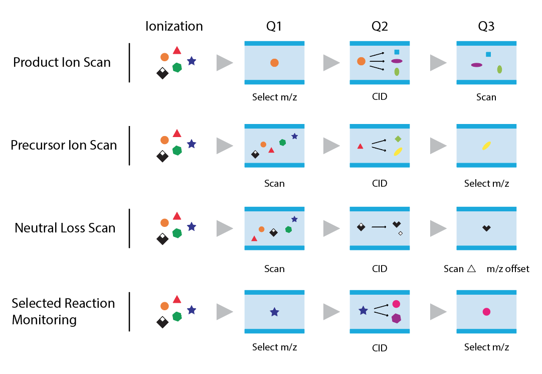
The TQMS consists of two quadrupole mass analyzers (Q1 and Q3) that are separated by a collision cell (q/Q2). While Q1 and Q3 are operated as mass analyzers to scan over a mass range or to monitor an ion of specific m/z value, the collision cell is used to fragment the precursor ions isolated in Q1 by subjecting them to high energy collisions with a neutral gas, such as argon, helium or nitrogen. It is possible to operate a TQMS in four different modes5 (Figure 3), namely:
- Precursor ion scan – the first quadrupole (Q1) is scanned over a mass range to select the precursor of a specific product ion (m/z value) which is then monitored in the last quadrupole (Q3).
- Product ion scan – Q1 is set to transmit only the pre-defined precursor (m/z) to the collision cell, while Q3 is scanned over a mass range to identify the fragments obtained under the experimental condition.
- Neutral loss (NL) – both Q1 and Q3 are scanned to identify all the precursors that give rise to the products by the loss of the same neutral (uncharged) species from all the precursors. The scan range of Q3 is offset by the NL value.
- Selected reaction monitoring (SRM) – both Q1 and Q3 are set to monitor specific m/z values for precursor and product ions. This mode is preferred for compound quantification due to its specificity and sensitivity. TQMS can be operated to monitor multiple precursor-to-product transitions of the same as well as different analytes.

Figure 3: Modes of operation of TQMS.
The fragmentation depends on the structure of the molecule and the experimental conditions, such as gas pressure and collision energy. Therefore, under a specific reaction condition, the fragmentation pattern is used along with the compound RT and its accurate mass value for identification. Moreover, monitoring of specific fragment ions helps to improve the sensitivity of detection and thus enables quantification of smaller amounts of the target compounds.
The QTOF mass spectrometer has a quadrupole mass analyzer and a time-of-flight mass analyzer separated by a collision cell. The quadrupole can be used either to transmit the ions or to isolate a specific precursor ion which is then fragmented in the collision cell. A small fraction of the ions are first pulsed into the TOF analyzer by a modulator and subsequently accelerated into the high-vacuum field-free region by applying high voltage. Ions with different m/z values travel at different velocities in the flight tube and are separated from one another. TOF mass analyzers offer high mass resolutions while being able to scan over large mass ranges quickly.
LC-MS analysis
LC-MS has been extensively applied for the analysis of both small molecules and large protein molecules in diverse matrices. Some examples of the applications of this technology are:
- quantification of genotoxic impurities in active pharmaceutical ingredients4
- detection of twelve model compounds that represent specific classes of doping agents, such as anabolic agents and simulants, in exhaled breath5
- quantification of drug metabolites in biological fluids
- detection of adulterants in food materials6 and dietary supplements7
- determination of alkylphenol ethoxylates (APEOs) in tannery sediments8
- quantification of personal care products in swimming pool and river water samples9
- quantification of nucleotides and their derivatives in bacterial cells10
- quantification of the proteome
- as a rapid assay for the detection of SARS-CoV-211
The technique has also been used in the analysis of drinking water, petrochemicals, soil, biopharmaceuticals, food and wine, and to detect per- and polyfluoroalkyl substances (PFAS) and pesticide residues.
Strengths and limitations of LC-MS
Strengths
LC-MS is suitable for the analysis of polar and non-polar compounds, as well as thermolabile molecules. These compounds can range from low molecular mass analytes with m/z values < 1000 Da, to very high molecular mass proteins with m/z values > 100,000 Da. The “soft ionization” of the compounds predominantly gives the molecular ion and the isotopic peaks which are helpful in the determination of the accurate mass and putative formula of the analyte. When coupled with the fragmentation spectra, it is possible to elucidate the structure of the analyte.
Although, typically a few milligrams of a pure compound are required for analysis by LC-MS, as little as 1 mg can be sufficient. By operating the MS in SIM or SRM mode, it is possible to achieve limits of detection in the range of ng/mL or even pg/mL. As only specific ions are monitored in SRM mode, selectivity is achieved even for analytes present in complex matrices.
Limitations
LC-MS instruments are expensive to own, operate and maintain. Expertise is required to run the instruments and analyze the data. The sample throughput is moderate in comparison to other analytical techniques. Since the spectra obtained depend on the experimental conditions, including the instrument type, the scope for compound identification via comparison to the reference spectra is limited. As a mass spectrometer is a destructive detector, care has to be taken when handling samples that may not be readily available or that are not obtainable in large amounts. As a laboratory-based rather than in-field technique, the analysis of unstable or reactive samples by LC-MS can prove challenging.
As only liquid samples can be injected into the column, solid samples have to be dissolved in a suitable solvent, or the analyte has to be extracted from the sample. Sample preparation by techniques such as liquid-liquid extraction (LLE) or solid-phase extraction (SPE) is essential for the extraction of target analytes from complex samples such as blood plasma, food and soil.12 This not only helps to improve the sensitivity of the analysis, but also reduces the contamination (discussed in the next section) of the system.
Common problems with LC-MS
While LC-MS confers several advantages for trace analysis in complex matrices, several precautions have to be taken to overcome the following challenges while using this technique.13
Contamination
The sensitivity, selectivity, reproducibility and resolution of analysis is impacted by contaminants, such as metal ions, phthalates, polyethylene glycol (PEG), slip agents, water and particles entering the system from various sources such as:
- reagents and solvents
- water used for preparing buffers
- chemicals leaching from glassware
- microcentrifuge tubes
- inlet filters
- solvent lines
- instrument parts, such as pump seals
- gases used for desolvation of the eluent in the source and in the collision cell
- the sample itself
The contaminants can interfere with the analysis by:
- suppressing or enhancing the ionization of analyte(s) in the source
- forming adducts with the analytes
- masking the analyte peaks and/or appearing as ghost peaks in the chromatogram
- making the baseline noisy
- fouling the system and the column, requiring frequent maintenance and replacement of parts
To minimize contamination:
- High-purity solvents, water and reagents should be used for the preparation of mobile phases.
- Freshly prepared mobile phases must be used to minimize the chance of microbial contamination of aqueous mobile phase and polymerization of acetonitrile (ACN).
- Use of soap or detergent to clean glassware should be avoided as they can be hard to remove and can cause interference during analysis.
- High purity gases (e.g., commonly used nitrogen gas of purity > 95%) must be used.
- Nitrogen generators must be well-maintained and the gas cylinder must be replaced when the pressure falls below the acceptable level.
- Analytes must be extracted from the sample matrix and chromatographic parameters optimized to improve the resolution of analyte peaks from interfering peaks.
Matrix effects
When analyzing biological samples, other sample constituents can suppress or enhance the ionization of the analyte in the source. To minimize the impact of the matrix, the analytes of interest should be isolated from it. Therefore, sample preparation is an important pre-requisite of LC-MS analysis. While this reduces matrix effects, it can be difficult to extract only the analyte(s) from the matrix. To prevent co-elution of interfering compounds, the chromatographic parameters can also be optimized. Preparation of standard solutions in analyte-free matrix (matrix matching) also helps to account for matrix effects. Known concentrations of isotopically-labeled internal standards, which experience similar ionization suppression or enhancement, are used to compensate for the matrix effects.
Carryover
Analyte peaks may appear in blank injections run after a high concentration sample as a result of sample carryover. This must be addressed by the use of cleaning protocols, such as repeated blank injections, needle washes and column conditioning, to ensure the sensitivity of the analysis is maintained.
Sample loss
Analytes, such as proteins and DNA, may be lost due to non-specific binding to laboratory consumables, such as the inner surface of microcentrifuge tubes. Adsorption of analytes impacts the accuracy and precision of the assay. Analyte loss can be minimized by using containers that have low surface adhesion. Another approach is to add blocking agents that minimize the interaction of the analyte with the inner surfaces of the containers.14
Mobile phase buffer selection
As the column eluent has to be removed prior to MS analysis, only volatile buffers, such as ammonium formate or ammonium acetate which will not precipitate in the source, can be used for the preparation of the mobile phases.
Maintenance
Regular maintenance of the mass spectrometer must be performed as per a pre-determined schedule to ensure the accuracy, reproducibility and trouble-free operation of the instrument and minimize unplanned downtime.
References
1. Dolan JW, Snyder LR. (1989) Injectors and autosamplers. In: Troubleshooting LC Systems. Humana Press, Totowa, NJ. doi:10.1007/978-1-59259-640-9_10
2. Medhe S. Mass spectrometry: Detectors review. Chem. Biomol. Eng. (2018); 3(4):51–58. doi:10.11648/j.cbe.20180304.11
3. Markgraf DF, Al-Hasani H, Lehr S. Lipidomics—Reshaping the analysis and perception of Type 2 diabetes. Int. J. Mol. Sci. 2016;17(11):1841. doi: 10.3390/ijms17111841
4. Chidella K, Dasari VB, and Anireddy J. Simultaneous and trace level quantification of five potential genotoxic impurities in ranolazine active pharmaceutical ingredient using LC-MS/MS. Am. J. Analyt. Chem, 2021;12:1–14. doi: 10.4236/ajac.2021.121001
5. Thevis M, Krug O, Geyer H, Schänzer W. Expanding analytical options in sports drug testing: Mass spectrometric detection of prohibited substances in exhaled breath. Rapid Commun. Mass Spectrom. 2017;31(15):1290–1296. doi: 10.1002/rcm.7903
6. . Development of a quantitative multi-compound method for the detection of 14 nitrogen-rich adulterants by LC-MS/MS in food materials, Food Addit. Contam: Part A. 2017;34(11):1842–1852. doi: 10.1080/19440049.2017.1372640
7. Dincel D, Olgan H, Canbaloğlu Z et al. Determination of dihydrocapsaicin adulteration in dietary supplements using LC-MS/MS. J. Chem. Metrol. 2020; 14(1):77–82. doi: 10.25135/jcm.36.20.01.1532
8. Mahalakshmi R, Pugazhendhi A, Brindhadevi K, Ramesh N. Analysis of Alkylphenol ethoxylates (APEOs) from tannery sediments using LC–MS and their environmental risks. Process Biochem. 2020;97:37–42, ISSN 1359–5113, doi: 10.1016/j.procbio.2020.06.015
9. , , , , . Determination of personal care products in water using UHPLC–MS after solid phase extraction with mesoporous silica‐based MCM‐41 functionalized with cyanopropyl groups. J. Sep. Sci. 2020;43:2142–2153. doi: 10.1002/jssc.201901148
10. Zborníková E, Knejzlík Z, Hauryliuk V, Krásný L, Rejman D. Analysis of nucleotide pools in bacteria using HPLC-MS in HILIC mode. Talanta. 2019; 205.120161. doi: 10.1016/j.talanta.2019.120161
11. Schuster O, Zvi A, Rosen O, et al. Specific and rapid SARS-CoV-2 identification based on LC-MS/MS analysis. ACS Omega 2021;6 (5):3525–3534. doi: 10.1021/acsomega.0c04691
12. Jickells S. (2021). Sample preparation. In Analytical Techniques in Forensic Science (eds R. Wolstenholme, S. Jickells and S. Forbes). https://doi.org/10.1002/9781119373421.ch4
13. Grebe SK, Singh RJ. LC-MS/MS in the clinical laboratory - Where to from here?. Clin. Biochem. Rev. 2011;32(1):5–31. PMID: 21451775
14. Silvester S, Zang F. Overcoming non-specific adsorption issues for AZD9164 in human urine samples: Consideration of bioanalytical and metabolite identification procedures, J. Chromatog. B, 2012;893–894:134–143. doi: 10.1016/j.jchromb.2012.03.004.

